Atomic force microscopy (AFM) is a nanoscale imaging technique that generates 3D topographical maps, enabling study of various surface properties and molecular structures.
Introduction to Atomic Force Microscopy (AFM)
Atomic force microscopy (AFM) is a powerful and versatile scanning probe microscopy technique that has revolutionized nanoscale imaging and measurement. Since its invention in 1986 by Binnig, Quate, and Gerber, AFM has become an essential tool for researchers in various fields, including physics, chemistry, biology, and materials science. This article will provide an overview of the basic principles and applications of AFM.
Principle of AFM
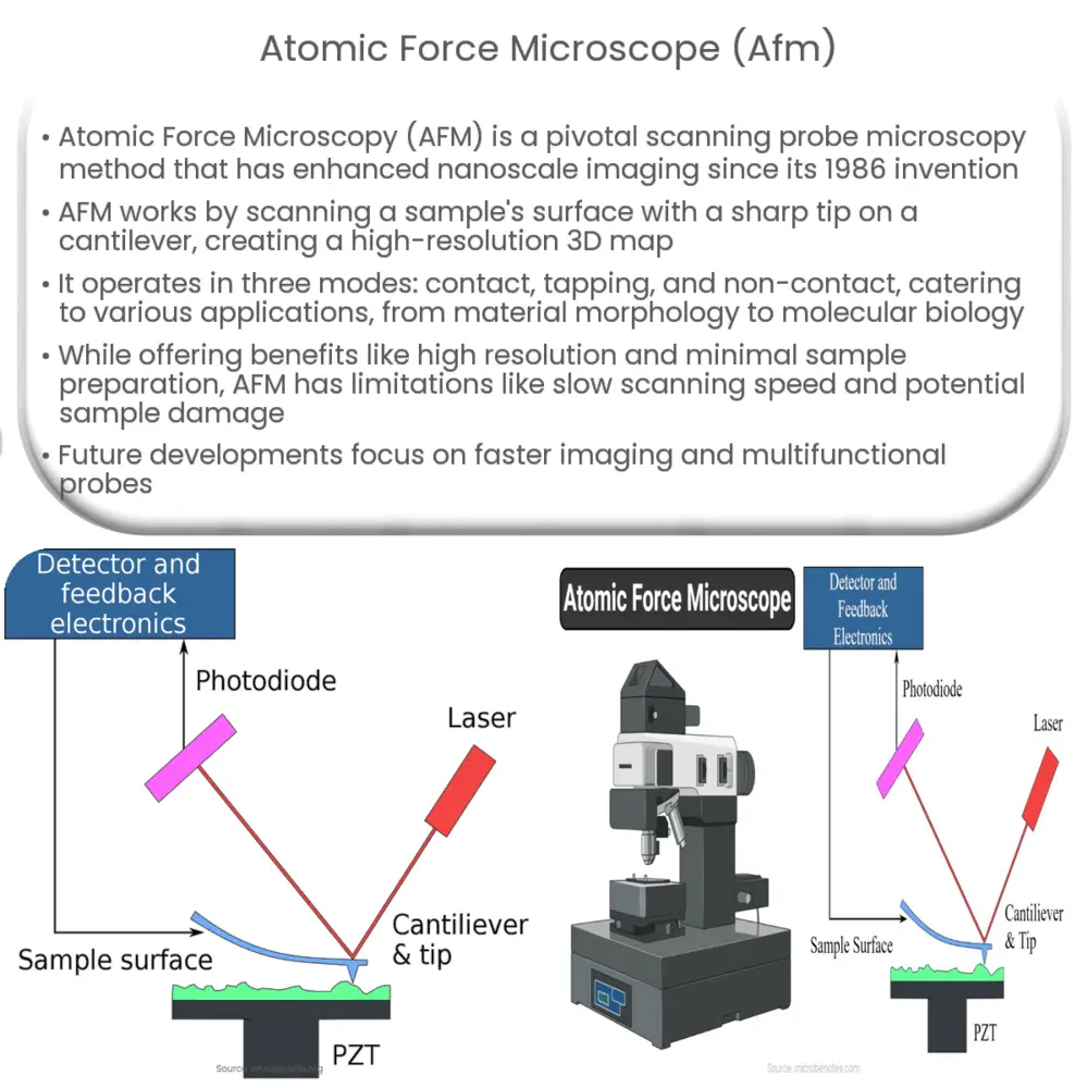
AFM operates by probing the surface of a sample with a sharp tip mounted on a flexible cantilever. As the tip scans across the sample surface, it interacts with the atoms and molecules on the surface, causing the cantilever to deflect. The deflection is monitored by a laser beam that is reflected off the cantilever’s backside and onto a position-sensitive photodetector. The amount of deflection is proportional to the force between the tip and the sample, allowing the AFM to generate a high-resolution, three-dimensional topographical map of the surface at the atomic scale.
Modes of Operation
There are three primary modes of operation for AFM: contact mode, tapping mode, and non-contact mode. Each mode provides different information about the sample and is suitable for specific applications.
Contact Mode
In contact mode, the tip is in continuous contact with the sample surface, and the force between the tip and the sample is kept constant by adjusting the height of the cantilever. This mode provides high-resolution topographical images and can be used to measure various surface properties, such as friction, adhesion, and electrical conductivity. However, the continuous contact between the tip and the sample can cause damage, especially for soft or fragile samples.
Tapping Mode
Tapping mode, also known as intermittent-contact or dynamic mode, involves oscillating the cantilever at or near its resonant frequency. The tip intermittently contacts the sample surface, reducing the lateral forces and minimizing damage to the sample. This mode is particularly useful for imaging soft, sticky, or loosely bound samples, such as biological specimens or polymer films.
Non-contact Mode
In non-contact mode, the tip is held a few nanometers above the sample surface, and the attractive forces between the tip and the sample are measured. This mode offers the least amount of sample damage and is suitable for imaging extremely delicate or reactive samples, but it generally provides lower resolution images compared to contact and tapping modes.
Applications of AFM
AFM has a wide range of applications in various fields, from studying the morphology and mechanical properties of materials to investigating biological processes at the molecular level. Some common applications include:
- Characterizing surface topography and roughness
- Measuring nanoscale mechanical properties, such as elasticity and adhesion
- Studying the structure and dynamics of biomolecules, such as proteins and DNA
- Investigating the electronic properties of materials, such as semiconductors and superconductors
- Examining the growth and assembly of nanoparticles and thin films
Advantages and Limitations of AFM
AFM offers several advantages over other microscopy techniques, including:
- High resolution: AFM can achieve atomic-level resolution in the x, y, and z axes, providing detailed, three-dimensional images of the sample surface.
- Minimal sample preparation: Unlike electron microscopy, AFM does not require conductive coatings, vacuum conditions, or other extensive sample preparations. This enables researchers to study a wide variety of samples, including insulators and biological specimens, in their natural state.
- Real-time imaging: AFM can provide real-time, in situ imaging of dynamic processes, such as molecular self-assembly, chemical reactions, or biological events.
- Simultaneous measurement of multiple properties: AFM can simultaneously measure various surface properties, such as topography, mechanical properties, and electrical conductivity, providing a comprehensive understanding of the sample.
However, there are some limitations to AFM:
- Slow scanning speed: AFM imaging can be time-consuming, especially for large areas or high-resolution scans.
- Limited field of view: The maximum scan range of AFM is typically limited to around 150 µm x 150 µm, making it unsuitable for analyzing large or macroscopic samples.
- Sample damage: While tapping and non-contact modes can minimize sample damage, the interaction between the tip and the sample may still cause alterations or deformations, especially for soft or delicate samples.
- Tip artifacts: The shape and condition of the AFM tip can influence the image quality and may introduce artifacts, requiring careful tip selection and calibration.
Future Developments in AFM
As AFM technology continues to evolve, researchers are developing new techniques and methodologies to address current limitations and expand the range of applications. Some promising areas of research include:
- High-speed AFM: Advances in scanner designs and control electronics have enabled high-speed AFM, allowing researchers to capture dynamic processes at the millisecond time scale.
- Multi-functional probes: By integrating additional sensors or functionalized tips, researchers can simultaneously measure multiple properties or perform specialized tasks, such as manipulating individual atoms or molecules.
- Correlative microscopy: Combining AFM with other microscopy techniques, such as electron or fluorescence microscopy, can provide complementary information and enhance the understanding of complex systems.
- Beyond imaging: AFM is increasingly being used for nanofabrication, enabling the manipulation and assembly of structures at the nanoscale for various applications, such as electronics, photonics, and biotechnology.
Conclusion
Atomic force microscopy is a powerful and versatile technique that has transformed our ability to visualize and manipulate the nanoscale world. With ongoing developments in instrumentation, probe design, and methodologies, AFM will continue to play a crucial role in advancing our understanding of materials, biological systems, and nanotechnology.
")
")